
Hydatidiform Mole: Symptoms, Causes, Treatment
What are the symptoms of a hydatidiform mole?
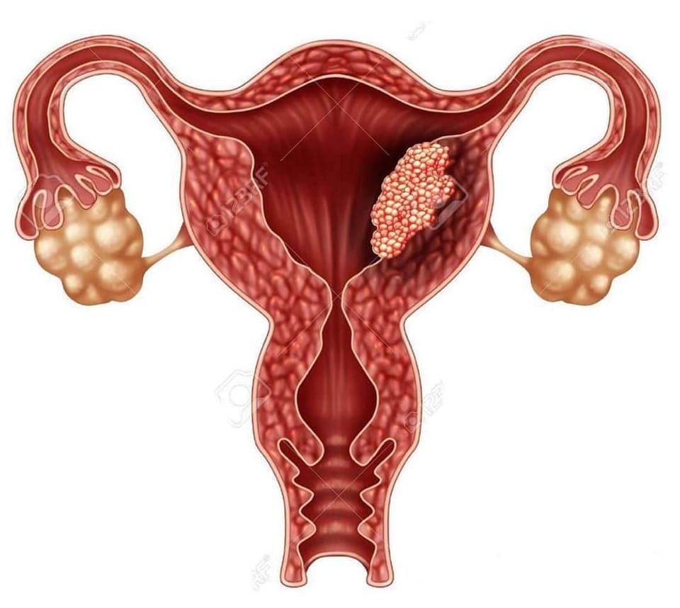
A hydatidiform mole, also known as a molar pregnancy, is an abnormal growth of tissue in the uterus that resembles a cluster of grapes. Symptoms of a hydatidiform mole can vary, but they may include:
- Vaginal bleeding: Vaginal bleeding, ranging from light spotting to heavy bleeding, is a common symptom of a hydatidiform mole. The bleeding may occur early in pregnancy and may be mistaken for a normal menstrual period.
- Grape-like clusters: In some cases, a hydatidiform mole may be detected during a pelvic exam as a mass in the uterus that resembles a cluster of grapes.
- Abdominal swelling: As the mole grows, it may cause the uterus to become enlarged, leading to abdominal swelling or bloating.
- Nausea and vomiting: Some women with a hydatidiform mole may experience nausea and vomiting, similar to morning sickness.
- High blood pressure: In rare cases, a hydatidiform mole may cause high blood pressure (gestational hypertension) or preeclampsia, a serious condition characterized by high blood pressure and organ damage.
- Hyperemesis gravidarum: Severe nausea and vomiting, known as hyperemesis gravidarum, can occur in some cases of hydatidiform mole.
- Anemia: Vaginal bleeding associated with a hydatidiform mole can lead to iron deficiency anemia, which can cause fatigue and weakness.
It’s important to note that many of these symptoms can also occur in a normal pregnancy or other conditions, so it’s important to see a healthcare provider for an accurate diagnosis if you experience any unusual symptoms during pregnancy. A hydatidiform mole is typically diagnosed through ultrasound and blood tests.
What are the causes of a hydatidiform mole?
A hydatidiform mole, also known as a molar pregnancy, is caused by an abnormal fertilization of an egg. There are two main types of hydatidiform mole:
- Complete hydatidiform mole: In a complete hydatidiform mole, an empty egg (ovum) is fertilized by a sperm, and the resulting mass of cells in the uterus has no fetal tissue. This occurs when the sperm fertilizes an egg that has lost its genetic material or when two sperm fertilize an egg.
- Partial hydatidiform mole: In a partial hydatidiform mole, an egg is fertilized by two sperm or by one sperm that duplicates its chromosomes. This results in an abnormal embryo with some fetal tissue, but it is not viable.
The exact cause of the abnormal fertilization in hydatidiform moles is not always known. However, risk factors for developing a hydatidiform mole include:
- Maternal age: Women over the age of 35 or under the age of 20 are at a higher risk.
- Previous molar pregnancy: Women who have had a molar pregnancy are at an increased risk of having another.
- History of miscarriage: Women who have had multiple miscarriages may be at a higher risk.
- Dietary factors: Diets low in animal fat and high in beta-carotene (found in fruits and vegetables) may be associated with an increased risk.
It’s important to note that most hydatidiform moles are benign (non-cancerous), but they can sometimes develop into a rare form of cancer known as gestational trophoblastic disease (GTD). Treatment for a hydatidiform mole typically involves removing the abnormal tissue from the uterus.
What is the treatment for a hydatidiform mole?
The treatment for a hydatidiform mole, also known as a molar pregnancy, typically involves removing the abnormal tissue from the uterus. Treatment options for a hydatidiform mole may include:
- Surgical removal: The most common treatment for a hydatidiform mole is a procedure called dilation and curettage (D&C). During a D&C, the cervix is dilated, and the abnormal tissue is gently scraped or suctioned out of the uterus.
- Monitoring: After a molar pregnancy is treated, regular monitoring is necessary to ensure that all of the abnormal tissue has been removed and to watch for any signs of complications or recurrence. This may involve regular blood tests to check the level of human chorionic gonadotropin (hCG), a hormone produced during pregnancy.
- Follow-up care: Women who have had a hydatidiform mole may be advised to wait for a period of time before trying to conceive again. They may also be advised to use contraception during this time to avoid a subsequent pregnancy.
- Treatment for gestational trophoblastic disease (GTD): In rare cases, a hydatidiform mole can develop into a form of cancer known as GTD. Treatment for GTD may involve chemotherapy or other treatments to destroy any remaining abnormal cells.
It’s important for women who have had a hydatidiform mole to receive follow-up care to monitor for any signs of complications or recurrence. Most women who have had a hydatidiform mole are able to go on to have successful pregnancies in the future, but close monitoring and care are important.


